Virus budding by cellular exocytosis
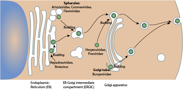
Budding at the endoplasmic reticulum (ER), ER -Golgi-Intermediate Compartment (ERGIC) or Golgi apparatus indicates that the viral particles are exported by cellular exocytosis.
There is a correlation between the site of budding and the site of replication.
Enveloped (+) RNA viruses and dsRNA viruses bud on ER or in the ERGIC, possibly because they replicate their genome in close proximity to these cellular organelles. Bunyaviruses bud at the Golgi, where they replicate in membrane invaginations.
Rotavirus genome replication and morphogenesis: role of the viroplasm
J. T. Patton, L. S. Silvestri, M. A. Tortorici, R. Vasquez-Del Carpio, Z. F. Taraporewala
Curr. Top. Microbiol. Immunol. 2006; 309: 169?187
The signal sequence of type II porcine reproductive and respiratory syndrome virus glycoprotein 3 is sufficient for endoplasmic reticulum retention
Do-Geun Kim, Chang-Seon Song, In-Soo Choi, Seung-Yong Park, Joong-Bok Lee, Sang-Soo Lee
J. Vet. Sci. 2013; 14: 307?313
Composition and three-dimensional architecture of the dengue virus replication and assembly sites
Sonja Welsch, Sven Miller, Ines Romero-Brey, Andreas Merz, Christopher K. E. Bleck, Paul Walther, Stephen D. Fuller, Claude Antony, Jacomine Krijnse-Locker, Ralf Bartenschlager
Cell Host Microbe April 23, 2009; 5: 365?375
Assembly and maturation of the flavivirus Kunjin virus appear to occur in the rough endoplasmic reticulum and along the secretory pathway, respectively
J. M. Mackenzie, E. G. Westaway
J. Virol. November 2001; 75: 10787?10799
Assembly and budding of a hepatitis B virus is mediated by a novel type of intracellular vesicles
Mouna Mhamdi, Anneke Funk, Heinz Hohenberg, Hans Will, H?seyin Sirma
Hepatology July 2007; 46: 95?106
Incorporation of spike and membrane glycoproteins into coronavirus virions
Makoto Ujike, Fumihiro Taguchi
Viruses April 2015; 7: 1700?1725
ER-Golgi intermediate compartment (ERGIC), also called The vesicular-tubular cluster (VTC)
endoplasmic reticulum (ER) or cisternae
Double membrane vesicle (DMV), tubes and spherules are membrane invagination in which viral replication takes place
The unique architecture of Bunyamwera virus factories around the Golgi complex
Juan Fontana, Noelia L?pez-Montero, Richard M. Elliott, Jos? Jes?s Fern?ndez, Cristina Risco
Cell. Microbiol. October 2008; 10: 2012?2028
Herpesvirus assembly: a tale of two membranes
Thomas C. Mettenleiter, Barbara G. Klupp, Harald Granzow
Curr. Opin. Microbiol. August 2006; 9: 423?429
The exit of vaccinia virus from infected cells
Geoffrey L. Smith, Mansun Law
Virus Res. December 2004; 106: 189?197