
Mastadenovirus (taxid:10509)
![]()
![]()
![]()
![]()
VIRION

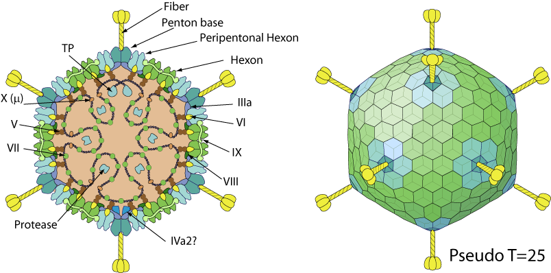
Non-enveloped capsid with a pseudo T=25 icosahedral symmetry  . The capsid diameter is about 90 nm. The capsid shell consists of 720 hexon subunits arranged as 240 trimers and 12 vertex penton capsomers each with a fiber protruding from the surface.

- Double jelly roll-fold major capsid protein [Hexon]
- Single jelly roll-fold penton capsid protein [Penton]
GENOME
 Monopartite, linear double-stranded DNA of 35-36kb encoding about 40 proteins. The genome has terminally redundant sequences which have inverted terminal repetitions (ITR). The terminal protein (TP) is covalently attached to each end of the genome.
GENE EXPRESSION
Transcription is nuclear, in three phases; early, intermediate (replication), and late (virion assembly). All genes are transcribed by host RNA pol II except virus-associated (VA) gene(s) of some primate adenoviruses transcribed by RNA pol III. Genes transcribed by RNA pol II give rise to multiple mRNA that are produced by alternative splicing and use of different poly(A) sites.
ENZYMES
- DNA-directed DNA polymerase B [POL]
- Protein-primed terminal transferase [POL on TP]
- Adenain (Peptidase C5) [Protease]
REPLICATION
NUCLEUS
- Attachment of the viral fibers to the host CAR adhesion receptor. Subsequent binding of the penton protein to host integrin entry receptors mediates internalization into the host cell by clathrin-mediated endocytosis of the virus and fiber shedding. Some serotypes also seem to use macropinocytosis.
- Disruption of host endosomal membrane by lytic protein VI releases the viral capsid in the cytosol.
- Microtubular transport toward nucleus of the viral genome still protected by the core protein VII and a partial capsid mainly composed of hexons and protein IX.
- Docking at the NPC and capsid disruption.
- Import of the viral genome into host nucleus mediated by core protein VII.
- Transcription of early genes (E genes) by host RNA pol II: these proteins optimize the cellular milieu for viral replication, and counteract a variety of antiviral defenses.
- Intermediate genes activate replication of the DNA genome by DNA strand displacement in the nucleus.
- Expression of L4-22K and L4-33K causes early to late switch. Transcription of late genes (L genes) by host RNA pol II, mostly encoding structural proteins.
- Host translation shutoff performed by the viral 100K protein.
- Assembly of new virions in the nucleus.
- Virions are released by lysis of the cell.
- Virion maturation by the viral protease.
Host-virus interaction
Adaptive immune response inhibition
The adenovirus E19 protein downregulates cell-surface expression of MHC-I by binding and retains the heavy chain in ER. E19 binds TAP and acts as a tapasin inhibitor, preventing class I/TAP association
.
Apoptosis modulation
Soon after infection, the expression of E1A protein drives the cell cycle into S-phase so that the virus can replicate its genome optimally . However, this arrest leads to the accumulation of host p53 protein and subsequently apoptosis. The apoptosis is then inhibited by different viral proteins including E1B 55K, E4 orf6 or the bcl-2 homolog E1B 19K .
Autophagy modulation
Autophagy is triggered after adenoviral infection and may facilitate viral replication and/or cell lysis. The E1B 19K viral protein interacts with host Beclin 1, removes Bcl-2 (a negative regulator of autophagy) from Beclin-1 and thereby promotes autophagy .
Cell-cycle modulation
The expression of E1A protein drives the cell cycle into S-phase so that the virus can replicate its genome optimally .
Innate immune response inhibition
Adenoviruses do not inhibit the cascade leading to production of interferon-beta directly. However, they evade the host innate immunity by targeting a cellular histone modification. Indeed, the E1A viral protein blocks the IFN-induced H2B mono-ubiquitination and thereby associated ISGs expression
.
Hongrong Liu, Lei Jin, Sok Boon S. Koh, Ivo Atanasov, Stan Schein, Lily Wu, Z. Hong Zhou
Science August 27, 2010; 329: 1038-1043
Carmen San Mart?n
Viruses May 2012; 4: 847-877
Matching UniProtKB/Swiss-Prot entries
(all links/actions below point to uniprot.org website)0 entry grouped by protein
Bat adenovirus 2 taxid:696069
Bat adenovirus TJM taxid:727977
Bat mastadenovirus WIV10 taxid:1788432
Bat mastadenovirus WIV11 taxid:1788433
Bat mastadenovirus WIV12 taxid:1788434
Bat mastadenovirus WIV9 taxid:1788436
Canine adenovirus serotype 1 taxid:10512
Canine adenovirus serotype 1 (strain RI261) taxid:69151
Canine mastadenovirus A taxid:10537
Chimpanzee adenovirus Y25 taxid:1123958
Equine adenovirus A serotype 1 taxid:46916
Equine adenovirus B serotype 2 taxid:67603
Human adenovirus 55 taxid:714978
| Protein | ModelArchive |
| L2 (Protein X) | ma-jd-viral-44608 |
| Protease (EC 3.4.22.39) (Adenain) (Adenovirus protease) (AVP) (Adenovirus proteinase)... | ma-jd-viral-13987 |
Human adenovirus A serotype 12 taxid:28282
Human adenovirus C serotype 2 taxid:10515
Human adenovirus C serotype 5 taxid:28285
| Protein | ModelArchive |
| 14.3 kDa protein (Control protein E4orf1) (E4 orf 1) | ma-jd-viral-04366 |
| 34 kDa protein (E4 34K) (E4 orf 6) | ma-jd-viral-05105 |